Preimplantation Genetic Testing (PGT)
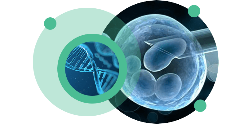
Preimplantation Genetic Testing, known by its English acronym PGT – Preimplantation Genetic Testing – is defined as a technique that allows the DNA of oocytes or embryos to be analyzed in order to detect genetic anomalies.
PGT encompasses three different types: PGT for detection of aneuploidies (PGT-A), for detection of monogenic diseases (PGT-M) and detection of structural genetic anomalies (PGT-SR).

About Preimplantation Genetic Testing (PGT)
Preimplantation Genetic Testing, known by its English acronym PGT – Preimplantation Genetic Testing – is defined as a technique that allows the analysis of the DNA of oocytes (polar blood cells) or embryos (in the cleavage or blastocyst phase) in order to detect genetic anomalies. Currently, with the evolution of techniques and conditions for embryo culture and embryo cryopreservation, the most common is for PGT to be performed on cells taken from the trophectoderm (outer layer) of the blastocyst, through a technique called embryo biopsy.
Complex and advanced genetic analysis techniques are used to perform PGT.
There are three types of genetic diagnosis:
- PGT-A – used to detect aneuploidies, i.e. the existence of extra chromosomes (trisomies) or fewer chromosomes (monosomies) in relation to the normal 46 chromosomes (e.g. Down syndrome)
- PGT-M – used to screen for monogenic diseases (caused by a single gene). This test aims to reduce the risk of transmitting a particular disease caused by mutations in a single gene (e.g. Cystic Fibrosis)
- PGT-SR – used to detect structural chromosomal abnormalities that can result in gain or loss of genetic material and which usually result in miscarriage.
Embryos with some type of genetic alteration may prevent a pregnancy from occurring or reaching term, or may give rise to a baby with a disease. The main objective of PGT is to increase the likelihood of pregnancy and reduce the risk of passing on genetic alterations or monogenic diseases to offspring.
Pre-implantation Genetic Testing is indicated in the following situations:
- Patients at risk of transmitting chromosomal abnormalities to their offspring
- Patients at risk of transmitting monogenic disease to their offspring
- Advanced maternal age (39 years or older)
- Successive implantation failures after IVF/ICSI cycles
- Patients with recurrent clinical miscarriage
- Previous pregnancy with genetic anomaly
Carrying out a treatment cycle using Pre-implantation Genetic Testing involves obtaining embryos through Medically Assisted Reproduction techniques, namely Intracytoplasmic Sperm Microinjection (ICSI).
In general terms, the process involves the following steps:
- ICSI treatment cycle – involves ovarian stimulation to develop several follicles simultaneously. These follicles are aspirated through a procedure called follicular puncture to collect the oocytes contained within them. In the laboratory, after processing the ejaculate sample and the oocytes, the microinjection technique is performed. The fertilized oocytes are kept in culture and will continue their development to obtain embryos (blastocysts) for 5 to 6 days.
- Embryo biopsy – procedure performed on embryos that develop normally and reach the blastocyst stage. In this procedure, some cells are removed from the embryo and sent for the desired genetic analysis, depending on the pathology or clinical case in question. The embryos subjected to biopsy are cryopreserved until the genetic results are obtained.
Embryo transfer and/or disposal – once the genetic results have been obtained, the embryos considered viable can be thawed and transferred to the patient’s uterus. Embryos with anomalies will be thawed and disposed of.
It is important to note that, as with all techniques, there is a percentage of error that can occur in PGT.
The error rate in PGT-M/SR is 1 to 5%, so the possibility of needing to carry out more tests during the pregnancy cannot be ruled out.
In the case of PGT-A, it is not possible to exclude the possibility that an embryo may be classified as transferable even if it contains an anomaly. This may happen because aneuploidy was not detected due to technical limitations or because the embryo is composed of both normal and aneuploid cells (mosaicism) but the cells analysed were all normal. Given these limitations, it is possible that embryos with aneuploidy may be transferred but, likewise, it is possible that embryos that would give rise to pregnancy and healthy babies may be discarded.
It is also important to note that the National Council for Medically Assisted Procreation (CNPMA) states that it has not been proven that carrying out PGT-A increases the success of ART techniques.
Given the complexity of this issue and the limitations associated with genetic diagnosis, it is extremely important that all these issues are discussed with your doctor.
Frequently Asked Questions
An embryo is made up of several cells and not all of them have the same genetic composition. An embryo with mosaicism has cells with normal chromosomal composition and cells with aneuploidy.
A monogenic disease is caused by mutations in a single gene. Examples of these diseases include Cystic Fibrosis, Fragile X Syndrome and Huntington’s Disease.
For a treatment cycle using PGT to be completed, each stage of the process must proceed normally, i.e., it must be possible to collect oocytes and sperm, fertilization must occur, and the fertilized oocytes must give rise to embryos (blastocysts) of sufficient quality to withstand embryo biopsy and freezing/thawing procedures.
Only embryos classified as transferable, without anomalies, may be transferred to the patient’s uterus. Non-viable embryos will be thawed and discarded.
