
Time-Lapse
Time-lapse technology applied to Clinical Embryology, namely in In Vitro Fertilization (IVF) and Intracytoplasmic Microinjection (ICSI) treatments, enables the capture and compression of thousands of images of the embryonic development process, generating an accelerated and fluid sequence of images in real time.
About Time-Lapse Technology
Time-lapse technology applied to Clinical Embryology, namely in In Vitro Fertilization (IVF) and Intracytoplasmic Microinjection (ICSI) treatments, enables the capture and compression of thousands of images of the embryonic development process, generating an accelerated and fluid sequence of images in real time.
It is very important to highlight that these images are of very high quality, which allows detailed monitoring of the embryos and consequently more precise assessments, such as detecting events that are rarely observed with conventional microscopy, such as asynchronous division or relevant morphological changes.
The application of this time-lapse technique to the domains of Clinical Embryology is particularly useful for obtaining more and better information about the development and quality of embryos, enabling the selection of those with the greatest implantation potential, contributing significantly to achieving better success rates.
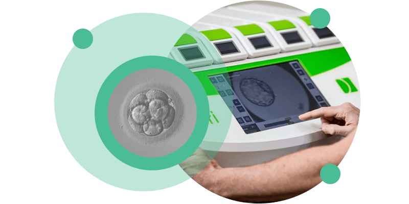
Time-lapse technology is incorporated into an embryo incubator that contains a built-in camera to capture photographs/images of embryo development at regular time intervals. The built-in camera is directly connected to a microscope.
The captured images are processed to form a time-lapse video. This way, the embryologist can evaluate in detail the different moments and stages of embryonic development.
CETI has the most advanced equipment with time-lapse technology, namely the Primo Vision™ Time-Lapse Embryo Monitoring System and the Geri® Time-lapse incubator.
The Geri Time-lapse incubator features a time-lapse system that monitors embryos from the oocyte to blastocyst stage (embryos on days 5/6 of development). The system captures an image of the embryo every five minutes, generating large amounts of relevant information so that embryologists can assess the cell division process without removing the embryos from the incubator.
In this way, embryos are objectively classified based on quality (high, medium, low), selecting the embryo most likely to reach the blastocyst stage and, therefore, the best embryo to transfer to the patient.
Unlike other incubators, Geri Plus has six independent spaces, each with its own high-resolution chamber. Each patient has their own incubator, ensuring that the embryos have personalized development conditions and their development is not affected by that of other embryos.
- Enhancement of results: since time-lapse technology aims to contribute to increasing success rates, since high-quality images allow detailed monitoring of embryos and more accurate assessments, such as detecting events that are rarely observed with conventional microscopy, such as asynchronous division or relevant morphological changes.
- Minimizing stress in embryo culture: the monitoring system placed inside the incubators prevents invasion of the embryos’ environment or their exposure to light.
- Use of embryo selection algorithms: these systems are based on selection algorithms tested on thousands of embryos and allow the best quality embryos to be optimally selected.
Frequently Asked Questions
Time-lapse technology is used at CETI in all In Vitro Fertilization (IVF) , Intracytoplasmic Microinjection (ICSI) and Cryopreserved Embryo Transfer treatments .
There is no additional cost. For CETI, incorporating and making advances in life sciences and technology available to customers is an integral part of its strategic pillar “Clinical Excellence Project”.
Time-lapse technology, by creating more and better information about the development and quality of embryos, increases the accuracy of selection, thus increasing the chances of success.