
CETI at the technological forefront
In vitro fertilization (IVF) has been one of the fastest developing fields of medicine and has been available for over four decades. The first successful birth following treatment was reported in 1978. Today, it is estimated that over 5 million children have been born as a result of IVF.
Despite technological advances in recent decades, the traditionally used method of morphological embryo selection was not sufficiently predictive to allow routine single embryo transfer. Therefore, new screening tools emerged, such as time-lapse technology.
CETI equipped with time-lapse equipment
CETI has the latest version of equipment based on time-lapse technology, the modern and complete Primo Vision™ Time-Lapse Embryo Monitoring System.
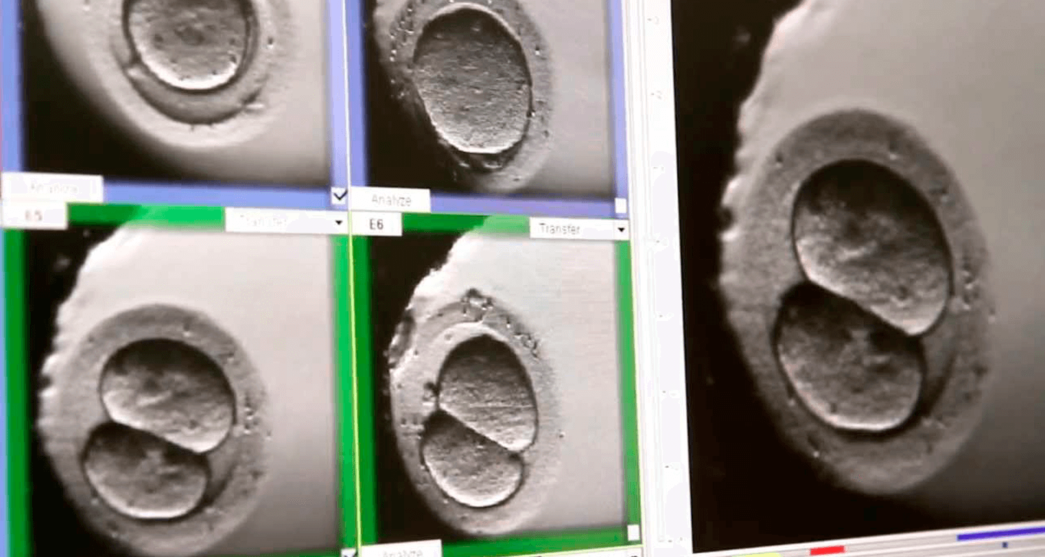
Time-lapse technology applied to Clinical Embryology consists of a non-invasive method of evaluating embryos, enabling the Embryologist to more carefully select the best embryos to be transferred to the mother’s uterus.
The time-lapse system enables continuous observation of embryos and, therefore, decision-making without disturbing the embryo culture, which can benefit their selection.
What does the time-lapse system consist of?
Primo Vision’s time-lapse system provides detailed insight into embryo development to improve selection and success rates.
Embryo monitoring with the time-lapse system allows continuous and non-invasive observation of the embryo without the need to remove the embryo from the culture conditions. The extra information about the cleavage pattern, morphological changes and embryonic development dynamics helps us to identify embryos with the greatest implantation potential.
These technological improvements allow us to objectively select embryos for transfer based on certain algorithms. Over the past 5-6 years, numerous studies have been published that confirm the safety of time-lapse technology.
What are the advantages of the time-lapse system:
- Enhancement of results: since time-lapse technology aims to contribute to increasing success rates, since high-quality images allow detailed monitoring of embryos and more accurate assessments, such as detecting events that are rarely observed with conventional microscopy, such as asynchronous division or relevant morphological changes.
- Minimizing stress in embryo culture: The monitoring system placed inside the incubators avoids unnecessary mobilization of embryos or their exposure to light.